NVIDIA’s research model combines a full-volume view with radiologist-style training, but it is not cleared for patient diagnosis.
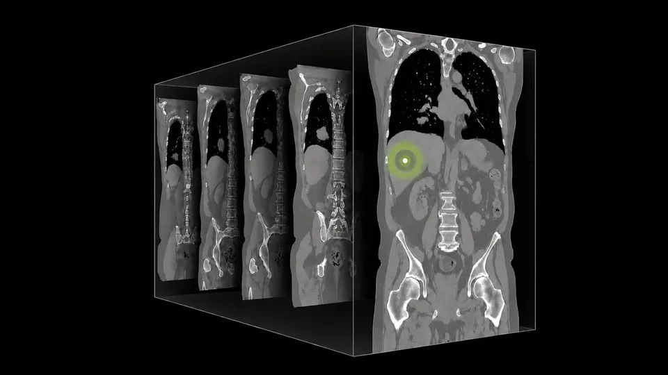
A computed tomography scan is not one picture. It is a stack of hundreds of thin images that together show the body in three dimensions.
NVIDIA says its new NV-Reason-CT model is designed to study that entire volume at once. It can produce structured reports, explain its findings in steps, and answer follow-up questions about possible diagnoses.
That matters because a slice-by-slice system can miss the way an abnormality changes across the scan. A mass, for example, may be defined by its shape, size, density and connection to nearby structures—not by what appears in one slice alone.
What changes when the model sees the whole volume?
Many medical vision systems begin with technology built for ordinary images. They process each CT slice as a separate two-dimensional frame, then try to combine the results.
NVIDIA says NV-Reason-CT instead uses a dedicated three-dimensional vision transformer. A vision transformer is a model that breaks an image into small pieces, called tokens, and learns how those pieces relate to one another.
For this model, those pieces are small cubes from the CT volume rather than flat image patches. NVIDIA says the system processes volumes resampled to 192 × 192 × 192 voxels, with each cube measuring 8 × 8 × 8 voxels. That creates a three-dimensional grid of 13,824 visual tokens.
The practical goal is to preserve continuity between slices. The model can consider whether a finding extends through several layers, how its shape changes, and where it sits relative to other anatomy.
This does not automatically make the interpretation correct. It gives the model a better representation of the information that radiologists already use when scrolling through a complete scan.
Why train it to explain its thinking?
A conventional medical AI system might return a label or probability: “possible lung nodule,” for example. That can be useful, but it gives a clinician little context about how the system reached the result.
NVIDIA trained NV-Reason-CT to produce radiologist-style reasoning. The model is taught to review anatomical regions, identify findings, consider alternatives and reach a structured conclusion.
It also supports multiple turns of conversation. A researcher or clinician could ask about a specific finding, request more detail about a possible alternative diagnosis, or ask the model to revisit part of its analysis.
The distinction is important: a conversational interface can make review easier, but fluent explanations are not proof that the underlying conclusion is right. A model can produce a convincing chain of reasoning while still overlooking an abnormality or misreading an image.
How does the training work?
NVIDIA describes a two-stage process.
First, supervised fine-tuning teaches the model from examples. These include structured reports, radiologist reasoning annotations and question-and-answer tasks about CT images. The curriculum contains approximately 550,000 structured question-and-answer examples covering chest and abdominal anatomy, localized findings, severity and abnormality detection.
The second stage uses reinforcement learning. In simple terms, the system receives feedback that rewards more accurate answers. NVIDIA says this feedback is anatomy-aware, so performance is judged within different body regions instead of relying only on one overall score.
The model is also trained to handle invalid questions and image-text pairs that do not match. That is intended to improve robustness, though it does not remove the need for testing on new hospitals, scanners and patient populations.
What results does NVIDIA report?
NVIDIA says NV-Reason-CT outperformed the published comparison systems it listed on the public CT-RATE benchmark. Its reported macro-F1 score was 0.614, compared with 0.581 for VoxelFM and 0.544 for Pillar-0. Macro-F1 is a way to average classification performance across multiple labels.
NVIDIA also reported an area-under-the-curve score of 0.871, slightly above VoxelFM’s 0.870. This score measures how well a system separates positive and negative cases across different decision thresholds.
Those numbers are useful for comparing the listed results, but they are not the same as proof of clinical benefit. The announcement does not establish prospective patient-care performance, error rates in routine use or independent replication of the results.
Is this something hospitals can use now?
NV-Reason-CT is an open research and development foundation, according to NVIDIA—not an autonomous diagnostic system or a cleared clinical product.
That means its immediate audience is researchers and developers. They could use the model and training approach as a starting point for experiments, then test and adapt it for a particular hospital’s scans, reporting process and patient population.
For engineers, the key change is architectural: native three-dimensional input avoids forcing a volume into unrelated two-dimensional pieces. For product teams, the more interesting feature may be the combination of structured reports and follow-up questions.
But deploying such a system would require more than downloading a model. Teams would need to measure its accuracy on external data, examine how often its explanations match the actual evidence, and determine its computing and latency requirements. Licensing and data-governance terms would also matter.
The useful next step is therefore not treating NV-Reason-CT as a replacement radiologist. It is testing whether full-volume processing improves specific reporting or review tasks enough to justify the added computing and validation work.